

Next: Shifts of the Spectra
Up: Data Reduction
Previous: Removal of the Photocathode
One drawback of magnetically focused image sensors is that they can have
geometric distortions if the electric and magnetic fields are not
absolutely uniform. This is evident in Fig. 8, where one
can see that the echelle orders are not perfectly straight. These
distortions must be calibrated and then eliminated if accurate
wavelengths are to be obtained. When confronting this problem, we
discovered a beneficial aspect of the disfigurement of the photocathode.
The apparent locations of the spots in our distorted electronic image
are very well defined by the sensitivity template; the spots have sharp
edges, and many of them have point-like centers that are even darker
than general (see Fig. 7). To find their true geometric
locations, we took a picture of the photocathode using a camera with a
long focus lens and densitometered the photograph. We then made point
by point comparisons of true and apparent locations over the whole field
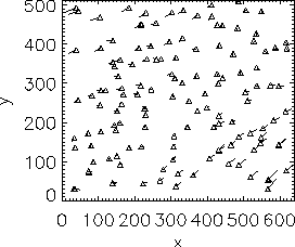
covered by the CCD. Fig. 9 shows the general
character and magnitude of the distortion (note the factor of 3
exaggeration of the offsets).
To remap an IMAPS exposure to a true coordinate system, we used the
information from the measured offsets shown in the figure. However, we
did not apply them directly![[*]](http://www.stsci.edu/icons//foot_motif.gif) , for to have done
so would have resulted in a crinkly mapping function that responded to
random measurement errors and the irregular sampling of points.
Instead, we evaluated
, for to have done
so would have resulted in a crinkly mapping function that responded to
random measurement errors and the irregular sampling of points.
Instead, we evaluated![[*]](http://www.stsci.edu/icons//foot_motif.gif) a best-fit 4th order polynomial that described the corrections
in x and y. Residuals between this fit and the measurements
appeared to be random, i.e., the polynomial appeared to respond to all
of the salient features of the distortion pattern, leaving only
measurement errors in the residuals.
a best-fit 4th order polynomial that described the corrections
in x and y. Residuals between this fit and the measurements
appeared to be random, i.e., the polynomial appeared to respond to all
of the salient features of the distortion pattern, leaving only
measurement errors in the residuals.
| Figure 9:
A map of the image distortions over the detector's format.
Flags show the differences between apparent positions in the electronic
image of the photocathode spots and their true locations, magnified by a
factor of 3 for clarity. |
 |
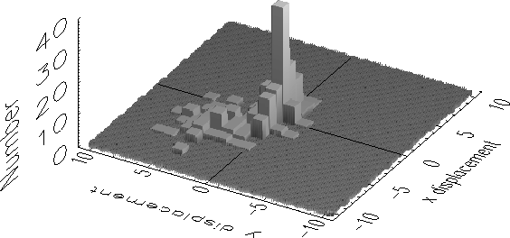
| Figure 10:
Numbers of various combinations of shifts in x and y seen
between successive exposures at a fixed echelle setting. Each unit of
the scale shown is in terms of the pixels in the image display used in
data reduction (equal to half of a CCD pixel). A large fraction of the
exposures showed little or no shift, as shown the tallest bars near the
origin. |
 |


Next: Shifts of the Spectra
Up: Data Reduction
Previous: Removal of the Photocathode
12/15/1998
![]() , for to have done
so would have resulted in a crinkly mapping function that responded to
random measurement errors and the irregular sampling of points.
Instead, we evaluated
, for to have done
so would have resulted in a crinkly mapping function that responded to
random measurement errors and the irregular sampling of points.
Instead, we evaluated![]() a best-fit 4th order polynomial that described the corrections
in x and y. Residuals between this fit and the measurements
appeared to be random, i.e., the polynomial appeared to respond to all
of the salient features of the distortion pattern, leaving only
measurement errors in the residuals.
a best-fit 4th order polynomial that described the corrections
in x and y. Residuals between this fit and the measurements
appeared to be random, i.e., the polynomial appeared to respond to all
of the salient features of the distortion pattern, leaving only
measurement errors in the residuals.